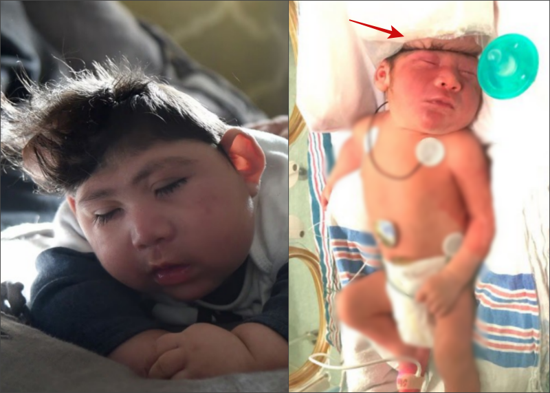
In a groundbreaking milestone for pediatric neurosurgery, seven-month-old Lucas—who was diagnosed in utero with exencephaly (a severe neural tube defect closely related to anencephaly)—is reaching developmental milestones, eating solid foods, and progressing through physical therapy alongside his family in New Jersey.
Diagnosed at 10 weeks of pregnancy, Lucas is believed to be among the first known infants to achieve long-term survival following a full-term exencephaly diagnosis, a condition traditionally considered universally fatal within hours of birth.
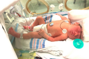
Understanding Exencephaly and Intrauterine Exposure
Exencephaly is an extremely rare congenital anomaly (occurring in roughly 3 out of 10,000 pregnancies) characterized by the complete or partial absence of the cranial vault (calvarium).
Because the skull fails to close during early embryonic development, the developing brain tissue remains directly exposed to amniotic fluid. Over the course of gestation, prolonged exposure to amniotic fluid and mechanical friction typically leads to severe tissue degradation (encephalomalacia), leaving most affected fetuses unable to survive long after delivery.
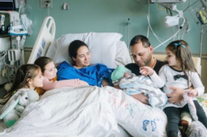
Lucas’s Clinical & Surgical Pathway:
- Fetal Diagnosis (10 Weeks) ──► Absence of cranial vault & exposed cerebral tissue
- Delivery & Initial Care ──► Born at term; sustained independent respiration and feeding
- Surgical Intervention ──► Resection of damaged sac & stabilization of basal ganglia
- 7-Month Milestones ──► Reaching typical developmental markers; physical therapy
Surgical Intervention and Neuroplasticity Strategy
When Lucas continued to breathe and feed independently hours after birth, Dr. Tim Vogel, Director of Pediatric Neurosurgery at the North Jersey Brain and Spine Center, formulated an innovative surgical plan to stabilize his exposed brain tissue and prevent fatal sac rupture or systemic infection.
The surgical strategy involved complex neurosurgical maneuvers:
-
Targeted Tissue Resection: Surgeons removed non-viable fluid-filled sacs and a portion of damaged subcortical tissue near the basal ganglia—the brain structure responsible for motor control and coordination.
-
Cranial Protection & Closure: Dr. Vogel protected the remaining functional brain tissue by constructing a protective skin barrier over the exposed defect to prevent fluid leaks and severe infection risks.
-
Leveraging Pediatric Neuroplasticity: Because an infant’s brain possesses extraordinary neuroplasticity—the ability to reorganize neural pathways—the surgical team anticipated that the remaining brain structures could adapt to manage motor and cognitive functions normally handled by the resected tissue.
Developmental Milestones and Ongoing Care
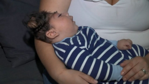
Seven months post-operation, Lucas is demonstrating developmental progress once thought impossible for his diagnosis:
-
Nutritional Progress: Transitioning successfully from liquid feeds to eating infant cereal and baby food.
-
Motor & Physical Therapy: Attempting to crawl and actively participating in early-intervention physical therapy sessions to build motor coordination.
-
Neurological Monitoring: Continuing long-term neurosurgical follow-up with Dr. Vogel to monitor intracranial stability and manage potential seizure risks.
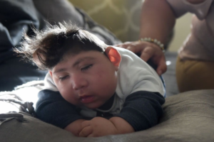
For his parents, Maria Santa Maria and Augusto, Lucas’s survival represents a profound medical triumph that offers new insights and hope to maternal-fetal care teams and families facing difficult prenatal diagnoses worldwide.